SweMagrose Protein G (1 mL)
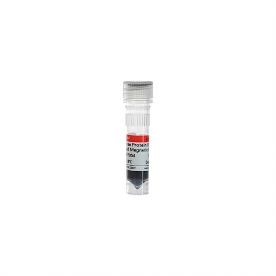
- Stock: In Stock
- Model: 0440
Product Description/Introduction
Protein G is an immunoglobulin-binding protein expressed by Streptococcal bacteria (C or G). Protein A is a cell wall surface protein found in Staphylococcus aureus with a molecular weight of 42kDa. Protein A and Protein G are functionally similar and can bind specifically to mammalian immunoglobulin (Ig). Recombinant Protein A and G with appropriate modifications bound to magnetic beads can be used for immunoprecipitation or antibody purification. while Protein G magnetic beads are suitable for the immunoprecipitation of human IgG1, IgG2, IgG3, IgG4, mouse IgG1, IgG2a, IgG2b, IgG3, rat IgG1, IgG2a, IgG2b, IgG2c polyclonal antibodies. Protein A magnetic beads are suitable for the immunoprecipitation of human IgG1, IgG2, IgG4, mouse IgG2a, IgG2b. (See Table I for specific information)
This product adopts the self-developed and produced ProteinG protein-labelled magnetic beads, compared with similar products in the market, this product binds antibodies more efficiently and has a lower non-specific binding rate, together with the optimized buffer, it can be convenient and efficient for immunoprecipitation experiments; it can be widely used in the isolation and purification of the target proteins in samples such as cell lysates, cellular secretion supernatants, serum, ascites, etc.
Product Information
Characteristics | Description |
Product content | 50 mg/ml magnetic beads in specific protective buffer |
Magnetization | Superparamagnetic |
Coupled protein | ProteinA |
M.W.of protein | ~25 kDa(ProteinA) |
Binding capacity | >1mg Mouse IgG per mL beads |
Specificity | antibodies from many different species, including mouse,human,rat,cow,goat and sheep |
Beads size | 30~150 μm |
Elution method | Acid or SDS-PAGE loading buffer elution. Note: If elute with SDS-PAGE loading buffer,the light(~25 kDa) and heavy(~50 kDa) chain of antibody will be denatured and release from the beads |
Application | IP,Co-IP,Protein purification |
Storage and Shipping Conditions
Ship with wet ice; Store at 4℃, valid for 12 months.
Product Components
Component | |||
SweMagrose ProteinG AntibodyPurifiedMagneticBeads | 1 mL | ||
Manual | 1pc | ||
Experiment preparation
Antibody purification related reagent formulations refer to the following, the user can be adjusted according to specific experimental conditions.
Component | Reagent combination |
Binding Wash Buffer | PBST:137 mM NaCl,2.7 mM KCl, 10 mM Na2HPO4, 2.0 mM KH2PO4, 0.1% Tween-20 |
Elution Buffer | 100 mM Gly, 0.1% Tween-20, pH 2.5 |
Neutralization Buffer | 1.0 M Tris-HCl, pH 9.0 |
Preservation Buffer | PBST,0.1%(v/v) Proclin 300 |
Manual procedure (purification of mouse ascites IgG as an example)
1. Sample processing:take 500 μL of ascites sample, add the binding wash buffer to make up 500 μL, if there are more protein precipitates in the sample, centrifuge the supernatant for experiments, which can improve the purity of the antibody.
2. Magnetic bead pretreatment: Vortex the antibody purified magnetic beads for 30 s to resuspend sufficiently, take 100 μL of 50% (v/v) SweMagrose Protein A Antibody Purified Magnetic Beads in another new 1.5 mL EP tube, magnetically aspirate it and discard the supernatant, wash it with 1 mL of binding washing buffer for twice, and then take the supernatant after magnetically aspirating.
Note:The amount of beads can be adjusted according to the amount of antibody in the sample.
3. Antibody adsorption: add the sample processed in step 1 to the magnetic beads in step 2, vortex and mix well, place the EP tube in a rotary mixer or manually turn the tube gently at room temperature (about 25°C) to make the magnetic beads full contact with the sample, turn it over for 15 min, and then place it on the magnetic separator rack for 30 s, and then discard the supernatant.
4. Magnetic bead washing:add 1 mL of binding wash buffer to the EP tube, resuspend with shaking and then magnetically absorb for 30 s. Discard the supernatant and repeat the operation 3 times.
5. Antibody elution:Add 0.5~1.0 mL of eluent to the EP tubes with the magnetic beads washed as described above, and resuspend the tubes rapidly by pipetting or vortexing, and then gently turn the tubes over in a turnover mixer or by hand at room temperature (about 25℃), and then separate the tubes by magnetic suction after turning over for 10 min, and then collect the supernatant into new EP tubes.
6. Antibody neutralisation:add a certain amount of neutralisation solution to the antibody eluate in step 5, generally 1/10 of the antibody elution volume (e.g., if the antibody eluate is 500 μL, the amount of neutralisation solution added is 50 μL), so that the pH value of the eluted antibody is maintained in a neutral environment, which is conducive to the maintenance of the biological activity of the antibody and the avoid antibody inactivation.
7. Post-treatment of magnetic beads: Wash the beads twice with elution solution after use, separate magnetically, anddiscard the supernatant; then wash three times with binding washing solution, separate magnetically, and discard the supernatant; resuspend the beads with 200 µL of preservation solution, and then store at 2~8℃.
Note
1. Please read these instructions carefully before proceeding with antibody purification.
2. Do not freeze or centrifuge the beads as this may cause irreversible aggregation of the beads.
3. Manual operation requires the use of a magnetic separation frame.
Table 1
Species |
Hygia SDS-PAGE Gel series are high performance precast polyacrylamide gels that are developed to separate a wide range of protein sizes by electrophor..
$87.00
Ex Tax:$87.00
Product Description/Introduction2 × Fast SYBR Green qPCR Master Mix (High ROX) is a special 2× premix for qPCR reaction using SYBR Green I chimeric fl..
$89.00
Ex Tax:$89.00
Product Description/Introduction2× Fast SYBR Green qPCR Master Mix (Low ROX) is a special 2× premix for qPCR reaction using SYBR Green I chimeric fluo..
$89.00
Ex Tax:$89.00
Product Description/Introduction2× Fast SYBR Green qPCR Master Mix (None ROX) is a special 2× premix for qPCR reaction using SYBR Green I chimeri..
$89.00
Ex Tax:$89.00
Product Description/Introduction2×SYBR Green qPCR Master Mix (High ROX) is a ready-to-use solution optimized for quantitative real-time PCR and t..
$89.00
Ex Tax:$89.00
Product Description/Introduction2×SYBR Green qPCR Master Mix (None ROX) is a special 2× premix for qPCR reaction using SYBR Green I chimeric fluoresce..
$89.00
Ex Tax:$89.00
Hygia SDS-PAGE Gel series are high performance precast polyacrylamide gels that are developed to separate a wide range of protein sizes by electrophor..
$87.00
Ex Tax:$87.00
0.9% NaCl..
$17.00
Ex Tax:$17.00
1000ml PES membrane Vacuum Filtration Systems With ISO9001&13485 certified, 1000ml PES membrane Vacuum Filtratio..
$243.00
Ex Tax:$243.00
Product Description/Introduction2 × Fast SYBR Green qPCR Master Mix (High ROX) is a special 2× premix for qPCR reaction using SYBR Green I chimeric fl..
$89.00
Ex Tax:$89.00
Product Description/Introduction2× Fast SYBR Green qPCR Master Mix (Low ROX) is a special 2× premix for qPCR reaction using SYBR Green I chimeric fluo..
$89.00
Ex Tax:$89.00
Product Description/Introduction2× Fast SYBR Green qPCR Master Mix (None ROX) is a special 2× premix for qPCR reaction using SYBR Green I chimeri..
$89.00
Ex Tax:$89.00
|







1-250x250.jpeg)
1-250x250.jpeg)


